How Do Cataracts Develop?
Age-related cataracts develop in two ways:
1. Clumps of protein reduce the sharpness of the image reaching the retina, resulting in a blurred image.The lens consists mostly of water and protein. When the protein clumps up, it clouds the lens and reduces the light that reaches the retina. The clouding may become severe enough to cause blurred vision. Most age-related cataracts develop from protein clumping. When a cataract is small, the cloudiness affects only a small part of the lens. You may not notice any changes in your vision. Cataracts tend to “grow” slowly, so vision gets worse gradually. Over time, the cloudy area in the lens may get larger, and the cataract may increase in size. Seeing may become more difficult. Your vision may get duller or blurrier.
2. The clear lens slowly changes to a yellowish/brownish color, adding a brownish tint to vision.As the clear lens slowly colors with age, your vision gradually may acquire a brownish shade. At first, the amount of tinting may be small and may not cause a vision problem. Over time, increased tinting may make it more difficult to read and perform other routine activities. This gradual change in the amount of tinting does not affect the sharpness of the image transmitted to the retina.If you have advanced lens discoloration, you may not be able to identify blues and purples. You may be wearing what you believe to be a pair of black socks, only to find out from friends that you are wearing a different color of socks.
Cataract Surgery
The lens is divided into three different portions: the center part called the nucleus, the outer part called the cortex, and surrounding both of these portions is a thin transparent membrane called the lens capsule which is like a  piece of saran wrap, or the outer skin of a sausage. The inner nuclear part is actually more dense than the outer, soft, cortical part. In order to remove a cataract we must first create a circular hole in the lens capsule and then take the inner nucleus of the lens out, which is soft like the inside of a sausage.
The inner nucleus must be removed with a phacoemulsification machine (phaco – lens; emulsification – to liquify). This machine has a metal probe that vibrates back and forth at a high frequency in order break into tiny pieces (emulsify) the central nucleus and gently sucks (aspirates) those pieces out of the eye. Once the central nucleus is removed, we then use other instruments to remove the softer cortex from the eye and try to polish up the capsule at the back, in order that as few cells as possible are left there. In most cases, the artificial lens (IOL) is then inserted to replace the focusing power that was lost with removal of the natural lens.
THE CATARACT SURGERY PROCEDURE
Cataract surgery is relatively simple and can be completed on an outpatient basis in about 20 minutes. The following five steps describe the process you will undergo when receiving cataract surgery:
PREPARING THE EYE
To ensure that cataract surgery is as comfortable as possible, two forms of anesthesia are used to numb your entire eye. Topical anesthesia bathes and numbs the surface of the eye, while the intraocular medication Xylocaine eliminates all sensation inside of the eye. Dr. James Gills developed this method of numbing the eye without needles in order to maximize patient comfort. It is now used by cataract surgeons throughout Florida and the world.
PREPARING THE SELF-SEALING INCISION
Once the eye is completely numb, an instrument is used to make a tiny, beveled “self-sealing” incision. This self-sealing incision allows the eye to heal without stitches. It works because the eye’s internal pressure holds the incision tightly closed. The self-sealing incision is less than 2.5 mm long, and is made at the edge of the clear cornea (the transparent covering of the front of the eye).
CATARACT REMOVAL
Cataracts form inside of the lens capsule, which is like an elastic bag that holds the lens in place. To remove the cataract, the front portion of the lens is opened. Next, a tool called a phacoemulsifier is inserted through the incision. The phacoemulsifier is used to gently break up the cataract with ultrasonic vibrations and then remove it out of the lens capsule.
IOLS REPLACE THE CLOUDY LENS
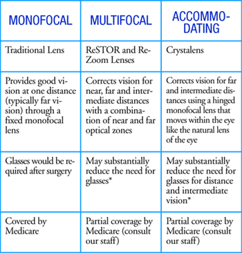
After the cataract has been completely removed, an intraocular lens (IOL) is inserted in place of the natural lens. This allows light to focus on the retina, resulting in clearer vision. There are many types of lenses that can be placed in the eye. IOLs can correct a wide range of preexisting refractive problems, including farsightedness, nearsightedness, and even astigmatism.
ANTIBIOTICS AND ANTI-INFLAMMATORY MEDICAIONS PLACED IN THE EYE
Before your surgery is complete, antibiotics and anti-inflammatory medicine will be placed inside your eye. The antibiotics will reduce your risk of infection to 1/20 the national average, while the anti-inflammatory medicine will promote healing and lessen your need for post-operative eye drops, which can be inconvenient and cause side effects.